Snake Eyes & Retained Eye Caps
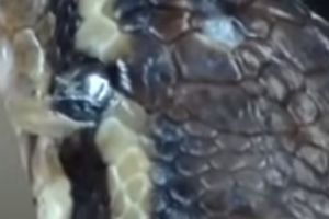
Snake eye caps are a transparent scale (actually part of the skin) and can have a number of potential issues.
Snakes evolved from lizards but have dramatically different eyes. These differences are cited widely as compelling evidence that snakes had fossorial and nocturnal ancestors.
[1] In fact, most scientists think that snakes had to somehow “reinvent their eyes”.
All snakes and some lizards have a fused, immobile lid covering the eyes, commonly referred to as eyecap (also known as the brille[2] or spectacle), but they do not have eyelids.
Because it is transparent, many think that a snake’s eyes are always open, when in fact, their eyes are always closed.
Even if under normal conditions the eye caps are clear and cannot be distinguished, they become cloudy when the animal is becoming ready for ecdysis. At that time then, eye caps become visible as a cover over the eye.
When snakes molt, the eye caps are also shed, generally inside out, as part of their skin.
The eye caps may be pathologically retained (for example if mites are present, with low humidity or if the eye is infected).
Retained eye caps have been associated with the development of corneal disease and panophthalmitis.
Snake Eye Facts
Fossorial lizards and mammals cluster together, whereas snakes are widely separated from these taxa and instead cluster with primitively aquatic vertebrates.
This indicates that the eyes of snakes most closely resemble those of aquatic vertebrates, and suggests that the early evolution of snakes occurred in aquatic environments. [3]
The shape of the pupil can give a clue to the diurnal or nocturnal activity patterns in a reptile. Some species have round pupils, others elliptical openings.
- Species with round pupils tend to be active during the day while those with slit pupils tend to be active at night.
- Coral snakes and all-new world non-venomous snakes (except the boa constrictor) have round pupils, while pit vipers have vertical slit pupils.
Snakes have adapted their vision to their activity period.
- Diurnal snakes have eye lenses that act as sunglasses, filtering out ultraviolet light and sharpening their vision
- Nocturnal snakes have lenses that allow ultraviolet light through, helping them to see in the dark.
The above means that some snakes can see other wavelengths, aside from visible light. This is extremely useful to detect prey in low light conditions, as they can perceive their body heat.
- For example, Pit vipers and some pythonomorphs (pythons and boas) can detect infrared radiation, being able to see the thermic signature around them.
The structure of the snakes eyes is mostly identical to that of the rest of the tetrapods.
A difference is a focusing method: while most tetrapods focus by changing the curvature of the crystalline lens, snakes focus moving the crystalline lens forward and backward.
Subterranean, more primitive snakes present quite simple eyes, with only rods that allow them to distinguish light and darkness. The sight is reduced, or even eyes are absent in some fossorial snakes. In general terms, snakes heavily rely on the other senses more than on their sight.
Snakes are dichromatic, meaning they can see two primary colors, blue and green, while humans are trichromatic: our cones react to wavelengths of light that allow us to see three primary colors, red, blue and green
Snakes Eye Anatomy
- The conjunctiva – is the tissue lining the inner lids to the sclera or eyeballs itself.
- The sclera – the sclera is composed entirely of tendinous connective tissue as the eye has no ossicles (unlike other reptiles)
- The cornea is the clear surface of the globe. Between the eye caps and the cornea, there are tear-like secretions.
- The retina – It is avascular and the membrane vasculosa retinae supply nutrients. The retina is usually grey mottled with white or red spots and appears with semi-opaque nerve fibers radiating uniformly outwards from the optic disc that is obscured in families having a conus.
- The iris is the colored portion inside the eye. By opening and closing, it allows light through the hole called the pupil onto the light receptors or retina on the back surface of the eye.
Diseases of the Eye in snakes
Snakes can be affected by several eye diseases, some specifically due to the fact they have eye caps.
As follows, we provide a list of the most common eye diseases in snakes that are either congenital or of environmental etiology:
1. Retained Shed on the spectacle
Many reasons may prevent proper shedding:
- a) inadequate humidity
- b) lack of suitable scabrous substrates upon which a snake can rub its chin and rostrum to initiate the molting process
- c) snake mites and ticks collecting along the spectacle margin
When this occurs you can try to gently remove the retained shed on the spectacle with a moistened cotton swat, or a little piece of cellophane tape.
To aid its removal, artificial tears (hypromellose) or hard contact lens wetting solution can be employed
However, if it does not come easily seek professional help, to avoid permanent damage. Although a partially lost spectacle will heal with successive ecdysis, exposure keratitis will develop if the entire spectacle is lost.
The below video shows the entire process of this.
2. Eye Trauma in snakes
Snakes might damage themselves from rubbing on the cage surfaces (abrasions, lacerations) or result damaged from bites from live prey.
They might heal on their own or with topical antibiotics; however, more severe lid lacerations would require surgical closure for proper healing.
Some wounds can be deep enough to destroy the globe, requiring enucleation or removal of the eye.
3. Corneal lesions in snakes
Corneal ulceration may be associated with foreign bodies or trauma as in other species.
Vets use a topical fluorescein dye and a black light to identify scratches or ulcers on the surface of the cornea.
Treatment for corneal damage is usually topical medication to prevent infection and promote healing. Occasionally, surgery is required.
4. Blockage of the nasolacrimal duct in snakes
Snakes produce tears to produce a region of lubrication between the cornea and the spectacle, allowing the free movement of the eye. If something is irritating the eye, there may be an overproduction of tears, or the tears may change consistency.
Tears cannot overflow the eyelids, as in mammals, so if a nasolacrimal duct is damaged there is a build-up of tears under the spectacle, which leads to a bullous spectaculopathy.
In humans and other species, the eye has a drainage system called the nasolacrimal duct that brings the tears to the nasal canal. If this duct is blocked, the tears have no place to go and will run down the face.
But, for species with a spectacle that develop a blocked nasolacrimal duct, the tears will build-up, thus creating a bulging spectacle.
The below video from Snake Discovery has a documentary of an eye removal by their vet which gives some insight into how the process would go.
The nasolacrimal duct can also be congenitally absent or become blocked by pressure from adjacent tissue (granulomas or neoplasia) or by fibrosis (burn injuries to the roof of the mouth). The fluid content may initially be clear but later become turbid and flocculent.
Although some of these blockages and infections will clear spontaneously, in many cases the infection is unrelenting and progresses to panophthalmitis or extends into the periocular tissue spaces.
Affected animals should be investigated for evidence of systemic infections.
Fluid can be drained through an incision in the ventral spectacle and antibiotics applied to the eye at this site.
Another approach is to create a new drainage route between the subspectacle space and the mouth (conjuntivoralostomy).
Systemic antibiotics are often required.
5. Cataracts in snakes
As humans, snakes can also suffer from cataracts.
With age, as well as with some systemic diseases, the lens (whose job is to focus the light beam on the light-sensing nerves of the retina) may become opaque, causing what we know as cataracts. This can lead to blindness.
However, as with humans, surgery is also available for snakes.
6. Conjunctivitis in snakes
Conjunctivitis is a term used for inflammation of the conjunctiva. Conjunctivitis in snakes may present itself as abscess formation under the spectacles.
Causes can be:
- a) poor water quality (that can lead to bacterial conjunctivitis)
- b) vitamin A deficiency
- c) trauma
As it usually takes a biopsy to tell the difference, it is advisable to treat for both with vitamin A supplementation and topical antibiotics. However, overdosing vitamin A can be toxic.
If there are sub spectacular abscesses, surgery will be required to remove the purulent material.
7. Uveitis in snakes
Uveitis is rarely diagnosed in reptiles although it occurs associated with systemic infectious disease, post hibernation disease (hyphema and hypopyon), trauma, and neoplasia.
The treatment utilizes topical (where applicable) and systemic antibiotic therapy and both steroid and non-steroidal anti-inflammatory drug therapy.
8. Microphthalmia in snakes
Microphthalmia is a congenital ocular malformation. Occasionally, snakes will be born with eye problems, the most common being this one. The eyes appear very small or nonexistent.
In the wild, these cases would carry a very poor prognosis of survival, but in captivity, many individuals do well.
9. Exophthalmia in snakes
In some snakes, eyes do not fit properly into the eye sockets. This is known as exophthalmia.
Congenital calcium deficiencies can lead to malformation of the skull, causing this bug-eyed look with the eyes, While the underlying disease can be treated, the skull deformity may be permanent.
Other causes for exophthalmia include retrobulbar abscesses, tumors, vascular obstruction, and edema. In these cases, the problem is not with the eye itself but with the structures around the eye that are causing it to be pushed outward.
Microphthalmos occur with some frequency in captive breed reptiles possibly as a consequence of inbreeding, or environmental conditions.
10. Enophthalmia in snakes
On the other hand, there is enophthalmia, which is sunken eyes. This can be a symptom of dehydration, weight loss, septicemia, or impending death.
A complete physical exam, as well as other diagnostic tests, may be necessary to determine the exact cause.
11. Other congenital abnormalities in snake eyes
Other congenital abnormalities in snake eyes include cyclopia and anophthalmos. These frequently occur with skeletal abnormalities.
Conclusion
Snake eye diseases are often identical to those found in mammals and birds. Although the approach to treatment is similar to that in mammals in many cases, anatomic differences in the snake eye and specifically the presence of eye caps in snakes present peculiar problems unique to these animals.
The first difference has to do with the fact eye caps appear impervious to topical medications, thus making treatment of the globe difficult. Eye caps are periodically replaced during molting cycles (ecdysis).
The second difference has to do with the fact ciliary muscles of reptiles are composed of striated muscle rather than smooth muscle fibers, and thus conventional mydriatic agents (parasympatholytics) such as tropicamide and atropine are ineffective in producing pupillary dilation.
In any case, some ophthalmological diseases in snakes can be treated at home, while in most cases, professional advice must be sought.
[1]Source: “The origin of snakes (Serpentes) as seen through eye anatomy”, by CAPRETTE, CH. Et al, in Biological Journal of the Linnean Society, Volume 81, Issue 4, April 2004, Pages 469–482, available at https://academic.oup.com/biolinnean/article/81/4/469/2643046
[2]Brille means “spectacles” or “glasses” in German, Norwegian and Danish and “shine” in French and Spanish.
[3]CARPETTE, CH. Et al, op. cit.
Table of Contents